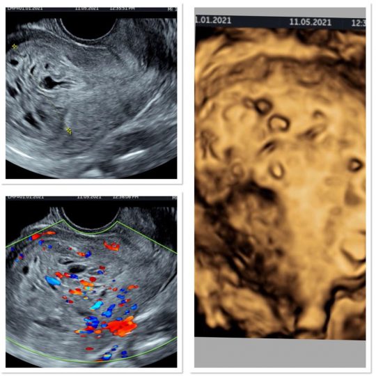
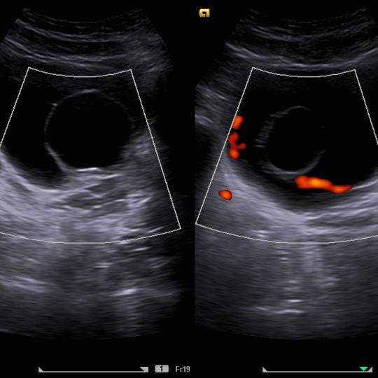
HYDATIDIFORM MOLAR PREGNANCY – A young 24years old lady presented with heavy bleeding with a positive UPT (urine pregnancy test). She was advised a transvaginal ultrasonography to confirm the status of pregnancy. The transvaginal ultrasound revealed extremly thickened and distended endometrial cavity with multiple cystic areas of varying sizes, giving a “cluster of grapes” appearance. No definte fetus was seen either within the cavity or outside. There was exaggerated vascularity on color doppler. In view of the clinical findings, positive UPT and markedly increased level of B HCG titres, a diagnosis of complete molar pregnancy was made.
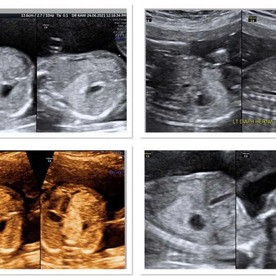
Diaphragmatic Hernia in Fetus – A 29 year old lady came for a scheduled Level II (anomaly )scan. The feuts showed normal growth but there was evidence of herniation of the abdominal bowel loops and occasionally stomach into the chest cavity. As a result, the heart was being pushed to the opposite side (ie the right). The residual lung volume on the right side was also being compromised.
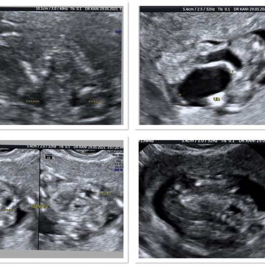
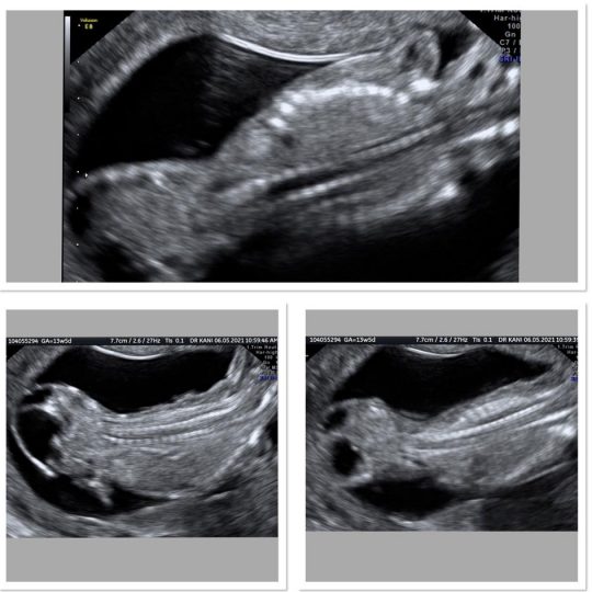
Multiple Defects at 13 weeks – A 30 year old lady came for a Level I (Nuchal) scan.The fetus was found to have multiple defects- altered spinal contour (exaggerated dorsal kyphosis), enlarged urinary bladder (megacystis) with back pressure changes to the kidneys and a low abdominal wall defect ( low examphalos) through which there was protrusion of the abdominal contents including the urinary bladder. The pregnancy was terminated.
INTUSSUSCEPTION – A 5 year old girl presented with history of a month long off and on pain abdomen and loose motions. She was advised an ultrasonography of the abdomen to find the cause. On ultrasonography, few mesenteric lymph nodes were seen. A thick- walled bowel loop segment with central hyperechoic core, giving a “target sign” appearance, was seen in the right flank, in the paraumbilical location. The anteroposterior diameter was approx 2.2cm. A diagnosis of Intussusception was given. Fortunately, the child had an uncomplicated course and was discharged.
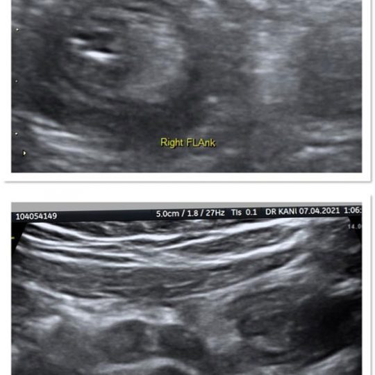
Sacrococcygeal Teratoma – A 30 year old lady in her 14 week of pregnancy presented for a Level I (Nuchal )Scan. A 2.2 x 2.2 x 2.1cm solid-cystic mass lesion was seen arising from the coccyx (tail bone) of the fetus . It was extending into the pelvis of the fetus. Rest of the struture of the fetus was alright.
Sacrococcygeal teratomas are sporadic in nature. They are of four types depending on the location. The ones arising in utero are associated with more complications.
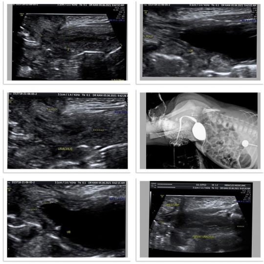
Patent Urachus – A month old new born presented with urinary discharge through the umbilicus (navel). Ultrasound revealed a 3.8 x 1 cm long fluid filled tract extending from the umbilicus to the dome of the urinary bladder. A contrast study was also done by injecting ~10ml of non ionic contrast into the navel. A tract was outlined upto the urinary bladder which filled up with contrast. The neonate was taken up for surgery and the tract excised.
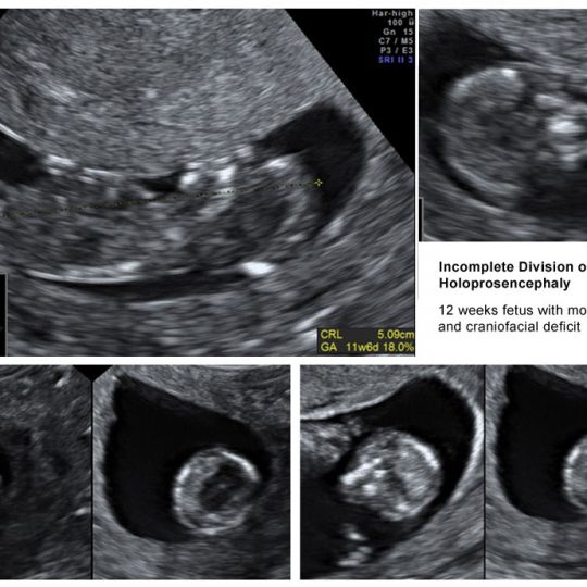
12 weeks fetus with monoventricle and craniofacial deficit
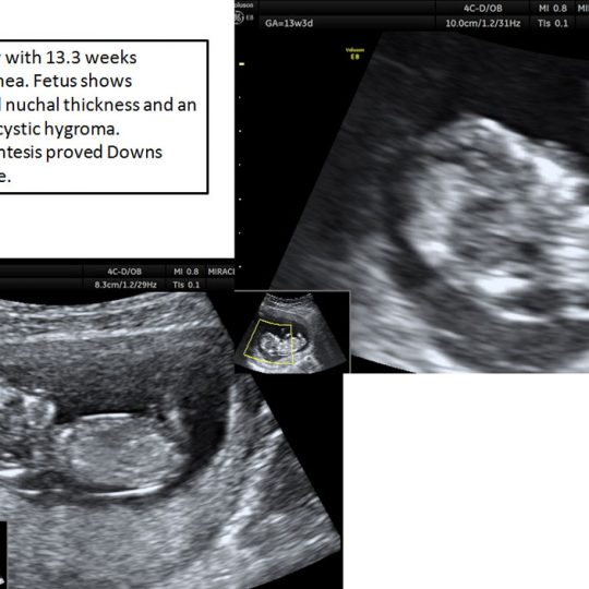
19 yr lady with 13.3 weeks amenorrhea. Fetus shows increased nuchal thickness and an evolving cystic hygroma. Amniocentesis proved Downs Syndrome.
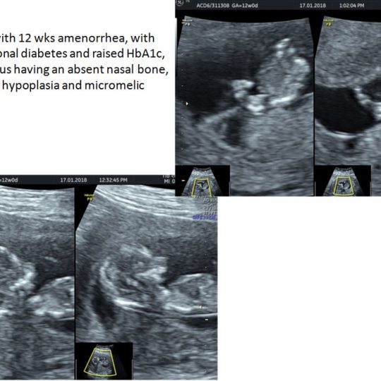
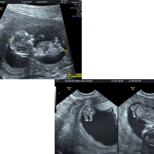
36 yr lady with 12 wks amenorrhea, with pre-gestational diabetes and raised HbA1c, with the fetus having an absent nasal bone, mandibular hypoplasia and micromelic limbs.
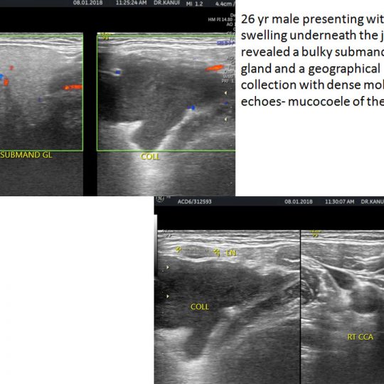
26 yr male presenting with a cystic swelling underneath the jaw. USG revealed a bulky submandibular gland and a geographical collection with dense mobile echoes- mucocoele of the duct.
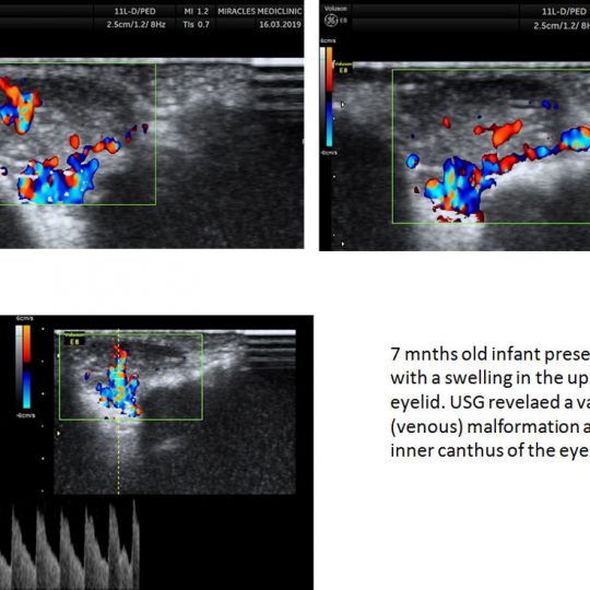
7 months old infant presented with a swelling in the upper eyelid. USG revealed a vascular (venous) malformation at the inner canthus of the eye.
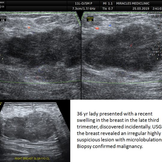
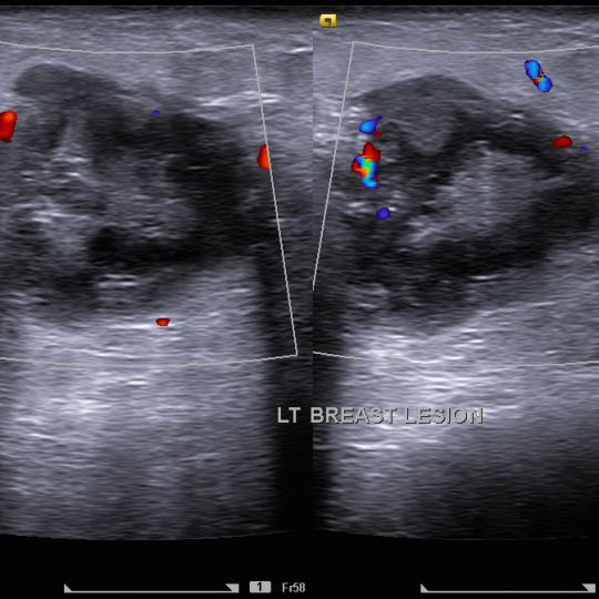
36 yr lady presented with a recent swelling in the breast in the late third trimester, discovered incidentally. USG of the breast revealed an irregular highly suspicious lesion with microlobulations. Biopsy confirmed malignancy.
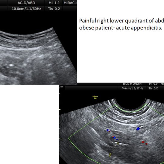
Painful right lower quadrant of abdomen in an obese patient- acute appendicitis.
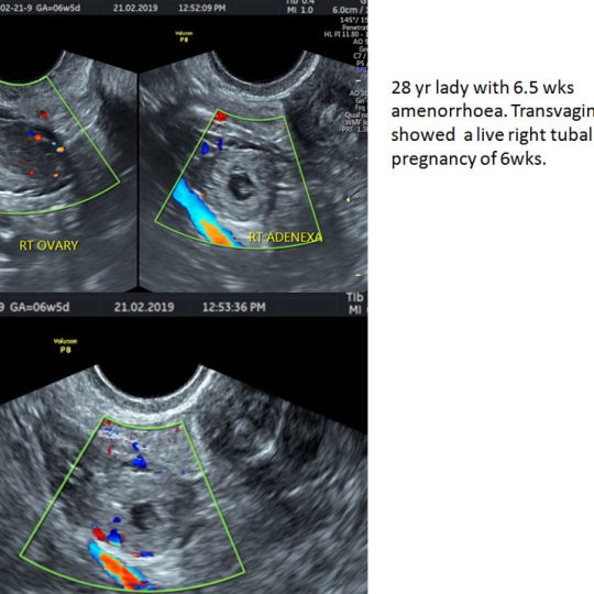
28 yr lady with 6.5 wks amenorrhoea. Transvaginal USG showed a live right tubal ectopic pregnancy of 6wks.
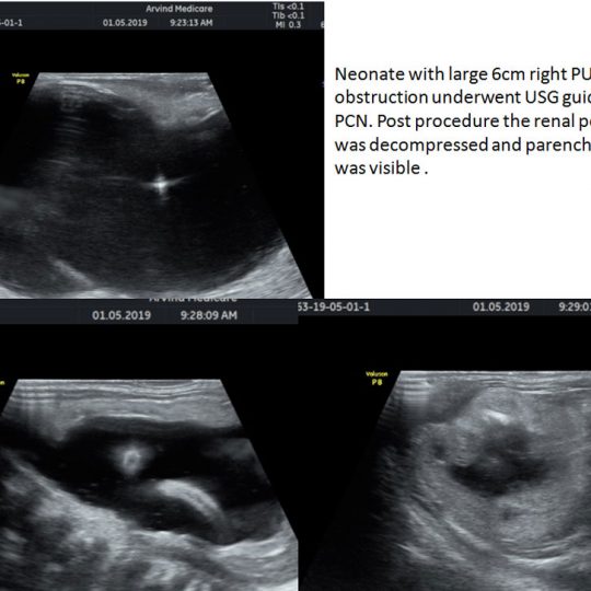
Neonate with large 6cm right PUJ obstruction underwent USG guided PCN. Post procedure the renal pelvis was decompressed and parenchyma was visible .
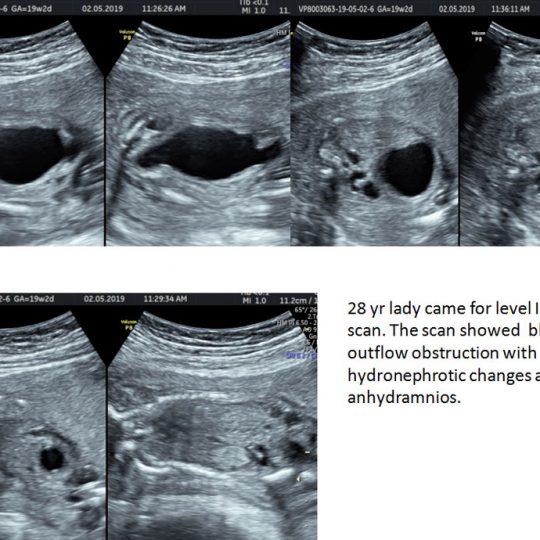
28 yr lady came for level II anomaly scan. The scan showed bladder outflow obstruction with hydronephrotic changes and anhydramnios.


