Calcified Abdominal Lesion and Increased Amniotic Fluid (Polyhydramnios)
Few months back, a pregnant lady of around 26weeks gestation walk in my clinic for an obstetric ultrasound, as she was feeling uncomfortable. I went through her previous records, which were all unremarkable and everything seemed to be going according to plan.
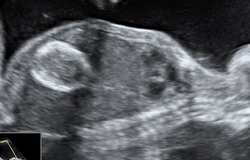
As I proceeded into the scan, I realised that the amniotic fluid was far more than the normal limit. Also, there was vague rim of calcification, below the left lobe of liver anteriorly. There was no ascitis or dilated bowel loops, and the stomach bubble was well dilineated. The growth of the fetus and the doppler blood flow were within normal range.
The lady carried on till 32 weeks but the amniotic fluid quantity didn’t come down, while her distress increased. She had a planned delivery and she delivered an otherwise healthy baby. An abdominal radiograph of the fetus was done which revealed an oblongish bubble of calcification below the hepatic shadow. The neonatal ultrasound revealed similar findings- a calcific rimmed lesion below the level of liver with mobile echoes within, with no other associated features. The pancreas was normal. My diagnosis was the possibility of a calcified pseudocyst on ultrasound.
The pediatric team did an exploratory laprotomy, which revealed a calcified meconium pseudocyst secondary to an in-utero sealed off perforated bowel. The baby did well post-op and is now an year old.