Please wait...


Blog

CLASSIC LIST

Scar-Pregnancy.jpg
20/May/2024

A 37 year old lady with a month old pregancy, came for a routine scan to confirm the status of the pregnancy. The first scan in a pregnancy is done to confirm the pregnacy, to localise the pregnacy (ie its placement- whether its is normally implanted or otherwise) , to check the age of the pregancy and to see if the cardiac activity of the embryo is recordable. On ultrasound , the pregnancy was seen to be within the uterus, but in the scar area of previous cesarian section. The upper cavity and cervical canal were empty. There was thinning of the scar myometrium. There was a fetus with near normal cardiac activity within the pregnancy gestational sac.

A cesarian scar pregnacy is very rare, though its incidence seems to be rising. It occurs due to poor healing of the cesarian scar tissue. If the pregnancy is allowed to progress, it may result in bleeding and even rupture of the uterus. This particular patient was managed by injecting potasium chloride into the growing fetus. Thus the fetal cardiac activity stopped. The pregnancy gestational sac gradually stopped growing too. Cesarian scar pregnancy cannot be terminated by the routine D and C (dilatation and curettage) method alone due to the risk of uterine rupture.

Caesarian Scar Pregnancy                   Scar-Pregnancy-3D


Calcified-Intrabdominal-Les.jpg
20/May/2024

Few months back, a pregnant lady of around 26weeks gestation walk in my clinic for an obstetric ultrasound, as she was feeling uncomfortable. I went through her previous records, which were all unremarkable and everything seemed to be going according to plan.

As I proceeded into the scan, I realised that the amniotic fluid was far more than the normal limit.  Also, there was vague rim of calcification, below the left lobe of liver anteriorly. There was no ascitis or dilated bowel loops, and the stomach bubble was well dilineated. The growth of the fetus  and the doppler blood flow were within normal range.

The lady carried on till 32 weeks but the amniotic fluid quantity didn’t come down, while her distress increased. She had a planned delivery and she delivered an otherwise healthy baby. An abdominal radiograph of the fetus was done which revealed an oblongish bubble of calcification below the hepatic shadow. The neonatal ultrasound revealed similar findings- a calcific rimmed lesion below the level of liver  with mobile echoes within, with no other associated features. The pancreas was normal. My diagnosis was the possibility of a calcified pseudocyst on ultrasound.

The pediatric team did an exploratory laprotomy, which revealed a calcified meconium pseudocyst secondary to an in-utero sealed off perforated bowel. The baby did well post-op and is now an year old.


Holoprosencephaly.jpg
20/May/2024

Case History

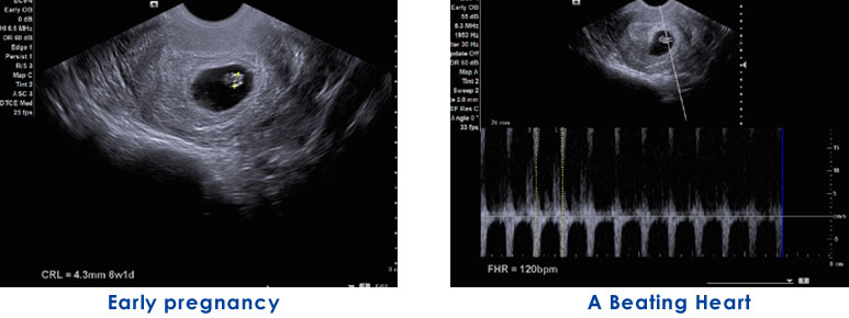
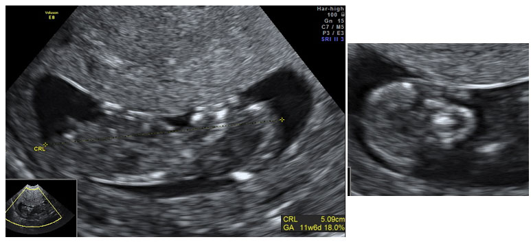
This is a case of a 32 years old lady, presenting at 12 weeks of gestation for a routine Level I scan (Nuchal Scan). Her previous scan at 8weeks showed a normal sized foetal pole with normal cardiac activity. She had a history of hypothyroidism for which she was on Tab Thyronorm 75/100mcg od. She had history of two previous miscarriages at 6-8weeks of gestation, first a chemical pregnancy and second an anembryonic pregnancy.

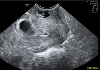
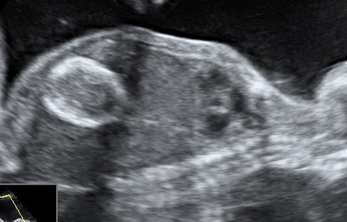
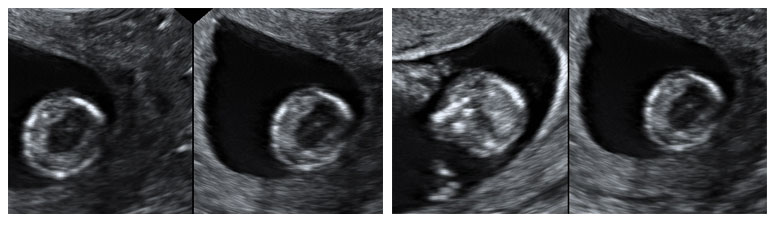
The present Level I scan revealed a small cranium with a monoventricle (absence of the butterfly appearance of chorod plexus). The cerebrum appeared to be fused anteriorly.  The posterior fossa  appeared normal for the gestational age.The orbital and nasal structures could not be delineated. Rest of the fetal body was unremarkable. The nuchal lucency was around 1.0mm. Ductus venosus showed normal forward flow.  My final diagnosis was of Holopresencephaly (semilobar type) with craniofacial abnormalities.

The lady presented a few days later with bleeding and signs of inevitable abortion.

Holoprosencephaly

Holoprosencephaly

Discussion

Holoprosencephaly refers to absent or incomplete division of the prosencephalon and is the most frequent malformation of the prosencephalon.  There is incomplete division of the prosencephalon during the fourth and eight week of gestation. Its incidence is 1 in 16,000 and 1 in 250 spontaneous abortion.

It is classified into 3 types according to the degree of cerebral involvement: alobar, semilobar and lobar. The clinical features vary depending upon the severity of holoprosencephaly.

  1. Alobar is the complete absence of division of the prosencephalon structures, resulting in complete absent interhemispheric fissure, corpus callosum, fused thalami and only oncerebral ventricle and facial dysmorphism. It is the most lethal.
  2. Semilobar, consisting in incomplete separation of cerebral hemispheres, connected in the frontal area, with a singular ventricular cavity and a partially fused thalami.
  3. Lobar, consisting of interhemispheric fissure, absent septum pellucidum and frontal horns communicate freely. Corpus callosum is normal or hypoplastic . It is the least severe form.

Another variant, the Middle Interhemispheric type has also been described.

The etiology of holoprosencephaly includes genetic and environmental factors. Environmental causes include maternal diabetes mellitus, alcoholism, inutero infections like CMV, rubella, toxoplasma, drugs like retinoic acid and cholesterol synthesis inhibitors. It can be transmitted in an autosomal dominant way. Mutation of SHH gene is the most frequent cause of familial holoprosencephaly. It is also asscoicated with Trisomy 13.

Prenatal diagnosis of holprosencphaly includes ultrasonography, fetal MRI, cytogentic analysis and molecular analysis of fetal DNA.

Prognosis is dependant on the degree of fusion and malformation of the brain as well as other health complication. Children born with lobar holoprosencephaly can survive for a few years but develop various neurological deficits.


DOPPLER.jpg
20/May/2024

One of the most anxiety filled examination in todays world is ‘follicular monitoring’ for wannabe parents, especially the lady. The desire to conceive, to hear the heartbeat of her baby and to finally hold one in her arms- is a long yet joyous journey.

One such special lady who had an earlier miscarriage was eagerly wanting to conceive. The previous miscarriage had shaken her but she did not want to give up the hope of raising a family. So after a few months of trying on their own, the folks were offered a chance to concieve with the aid of sonologically monitoring the ovulation. The first month was a simple baseline study. However amidst all hope there was premature ovulation of the growing follicle and it was in a sense a wasted cycle. Not one to give up she consulted a gynaecologist and along with folic acid she was put on DHEA as her AMH count was on the lower side.

Follicular-Monitoring

I started monitoring her on day 2/3 of her cycle to assess her ovaries for cysts and the endometrial ling of her uterine cavity. Fortunately there were no cysts. She was called back on the 8th day. There was a dominant follicle in her right ovary. She was called every alternate day till day 14 of her cycle, to monitor the size of the dominant follicle and the endometrial lining. Fortunately and God willing the follicle had rupture ( ovulated). The follicular study was complete. But another wait started.

The past keeps haunting till you let it go. It takes a lot of endurance and perseverance to escape from the tight hold of a past event gone bad. The next three to four week period of anticipation and apprehension must have been gruelling for her.

She missed her period in the next cycle and she came in at around 6 weeks for a scan. The feeling is akin to that of when one has take an important exam in one s life and the result is eagerly awaited. Presto! There in her womb was a little life beating its heart. There were tears of joys, sigh of relief and a round of congratulations.
The second phase of the journey to mother and parenthood commenced.

WHAT IS FOLLICULAR MONITORING
It is simply tracking the size of an active (dominant) follicle in the ovaries that contains an egg.
It is useful for accurately predicting when ovulation might be expected to enhance the chance of fertilisation and conception.

FOLLICULAR MONITORING

HOW IS FOLLICULAR MONITORING DONE?
It is usually done via transvaginal route (TVS) from the 8th day of cycle. The follicle is monitored till it reaches 22-24mm diameter and till it is is subsequently released from the ovary.

WHAT ARE THE BENEFITS?
It aids a couple in planning the best possible time to get pregnant. It also aids in planning an assisted reproductive technique- IUI (intrauterine insemination) and IVF (in vitro fertillization).


© Copyright Dr. Kanupriya 2023. All rights reserved.